Spine surgery has a reputation problem in PCMC. Patients picture a long incision down the back, hours in the operating room, weeks in hospital, months of recovery. That picture has been outdated for over a decade. Endoscopic spine surgery represents the most modern end of that evolution - an 8 millimetre incision, a thin tube, a high-definition camera and a procedure that often allows the patient to walk out of the hospital the same evening.
Dr. Swaroop Solunke offers endoscopic spine surgery for selected cases at his Wakad partner hospitals. The technique is excellent in the right patient. It is not appropriate for every spine problem. The first job is honest assessment of who qualifies and who does not.
What Is Endoscopic Spine Surgery?
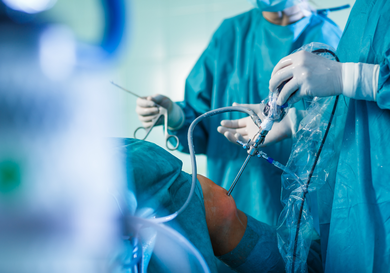
Endoscopic spine surgery is a minimally invasive technique where a 6 to 8 millimetre tube containing a high-definition camera and working channel is inserted through a small skin incision. The surgeon watches a live video feed of the spine on a monitor and uses tiny instruments through the same tube to remove the disc fragment, decompress the nerve, or address other pathology.
This is fundamentally different from open spine surgery (long incision, muscle stripping) and from microscopic spine surgery (smaller incision but still through muscle). Endoscopic technique works through natural anatomical corridors, often without cutting any muscle at all.
.png)
Two Main Endoscopic Approaches
Transforaminal Endoscopic Spine Surgery (TESSYS)
The endoscope enters from the side of the spine through the natural opening (foramen) where the nerve exits. The disc is approached from the side. Suitable for disc herniations that lie laterally or to the side.
Interlaminar Endoscopic Spine Surgery
The endoscope enters from the back, between two vertebral arches (laminae). Better suited for disc herniations that lie in the central or paracentral position.
Cervical Endoscopic Spine Surgery
Specialized endoscopic procedures for selected cervical (neck) disc problems. Less commonly done than lumbar endoscopic surgery.
Conditions Treated With Endoscopic Spine Surgery
Lumbar Disc Herniation (Slipped Disc) Causing Sciatica
The most common indication. The endoscope removes the herniated portion of the disc that is pressing on the nerve. Pain relief is often immediate.
Foraminal and Lateral Disc Herniations
Disc fragments that have migrated to the side, where they are technically harder to reach with traditional surgery. Endoscopic transforaminal approach is particularly suited for these.
Selected Cases of Foraminal Stenosis
Narrowing of the nerve exit foramen. Endoscopic foraminoplasty enlarges the opening to relieve nerve compression.
Recurrent Disc Herniations
Disc herniations that have come back after a previous discectomy. Endoscopic surgery avoids the scar tissue from the previous open surgery.
Selected Cases of Synovial Cysts
Fluid-filled cysts arising from spinal facet joints that compress nerves. Can be removed endoscopically in selected cases.
Strict Candidate Selection - Who Qualifies?
You May Be a Candidate If:
- You have a clearly identified disc herniation on MRI causing sciatica or arm pain
- Conservative treatment (physiotherapy, medication, injections) has failed for 6 to 12 weeks
- Your symptoms match the imaging findings precisely
- The disc herniation is in a position the endoscope can reach (most lumbar levels qualify)
- You do not have severe spinal instability
- BMI is below 35 (technically more difficult above this)
.png)
Endoscopic Surgery Is Not Suitable If:
- There is significant spinal instability requiring fusion
- There is severe central canal stenosis with major nerve compression
- There is significant deformity (scoliosis, severe spondylolisthesis)
- Multiple levels need extensive decompression
- Spine infection or tumour
- Anatomical features make endoscopic access technically impossible
Roughly 30 to 40 percent of patients who would be considered for traditional discectomy are also good candidates for endoscopic surgery. Dr. Solunke evaluates each patient individually with MRI, CT and clinical examination.
Advantages of Endoscopic Spine Surgery
Tiny Incision
8 millimetre incision versus 2 to 3 centimetres for microdiscectomy and 8 to 10 centimetres for traditional open discectomy. Cosmetic outcome is excellent.
Muscle-Sparing
The endoscope works through natural anatomical corridors without cutting muscle. This is why post-operative pain is dramatically less.
Local Anesthesia Possible
Many endoscopic spine surgeries can be done under local anesthesia with light sedation, especially in elderly or medically frail patients who cannot tolerate general anesthesia.
Fast Recovery
Most patients walk within 2 to 4 hours of surgery. Discharge on the same day or the next morning is standard. Return to desk work in 1 to 2 weeks.
Less Blood Loss
Average blood loss is under 50 ml - less than what is lost during a routine blood test.
Lower Infection Risk
Smaller wound, less tissue exposure, shorter operative time all contribute to a very low infection rate.
Preserves Future Options
If symptoms recur, traditional surgery remains available because endoscopic surgery does not produce the scar tissue that limits revision options.
How Endoscopic Spine Surgery Is Performed
Step 1 - Anesthesia. Local anesthesia with sedation, or general anesthesia depending on patient preference and surgical complexity.
Step 2 - Positioning. Most lumbar endoscopic procedures are done with the patient lying on the side or face down on a special table.
Step 3 - Skin incision. An 8 millimetre incision is made over the planned entry point, identified by fluoroscopy.
Step 4 - Endoscope insertion. A guide wire is placed first, followed by progressive dilators and finally the working tube and endoscope. Live video feed appears on a high-definition monitor.
Step 5 - Disc removal. The herniated portion of the disc is identified and removed using small specialized instruments through the working channel of the endoscope. Decompression of the nerve is verified.
Step 6 - Closure. The endoscope is removed. The 8 mm incision is closed with one or two stitches or simple adhesive strips.
Total surgery time: 30 to 90 minutes depending on the procedure.
Pricing for Endoscopic Spine Surgery in Pune
- Endoscopic discectomy (single level): Rs. 2 to 3.2 lakh
- Endoscopic foraminoplasty: Rs. 2.2 to 3.5 lakh
- Endoscopic decompression for stenosis: Rs. 2.5 to 4 lakh
- Cervical endoscopic procedures: Rs. 2.5 to 4 lakh
- Hospital stay: same-day or 24-hour stay in most cases
Most insurance plans cover endoscopic spine surgery on cashless basis. The cost difference between endoscopic and traditional microdiscectomy is generally moderate.
Recovery After Endoscopic Spine Surgery
Day of Surgery
Walking within 2 to 4 hours of surgery. Most patients walk to the bathroom independently the same evening. Discharge same day or next morning.
Week 1
Walking at home, gradual increase in activity. No heavy lifting (above 2 to 3 kg). Avoid prolonged sitting initially. Stitches removed at day 7 to 10.
Week 2 to 4
Most desk-job patients return to work. Light physiotherapy and graduated walking programme.
Week 4 to 8
Structured strengthening physiotherapy. Return to gym basics. Most patients return to all desired activities.
Month 3
Full functional recovery for the majority of patients. Heavy lifting and contact sports gradually resumed.