Knee arthroscopy is the everyday workhorse of modern knee surgery. The same camera-and-instruments approach that revolutionized orthopedics in the 1990s now treats most non-arthritic knee problems through three small incisions. Patients walk in, have surgery and often go home the same evening with a few stitches.
Dr. Swaroop Solunke performs knee arthroscopy at his Wakad clinic and partner hospitals in PCMC. The clinic handles a steady volume of meniscus tears, cartilage problems and patellar issues - particularly from sports injuries and wear-related problems in middle-aged patients.
What Is Knee Arthroscopy?
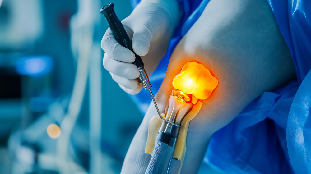
Knee arthroscopy is a minimally invasive surgical procedure where a thin tube with a camera at its tip (the arthroscope) is inserted through a small incision in the skin. The camera sends a magnified image to a high-definition monitor. Through one or two more small incisions, specialized instruments are inserted to repair, trim, reattach or reconstruct tissue inside the knee.
The incisions are 5 to 8 millimetres each. There is no large open scar. Soft tissue trauma is minimal. Hospital stay is short - most patients are discharged the same evening or the next morning.
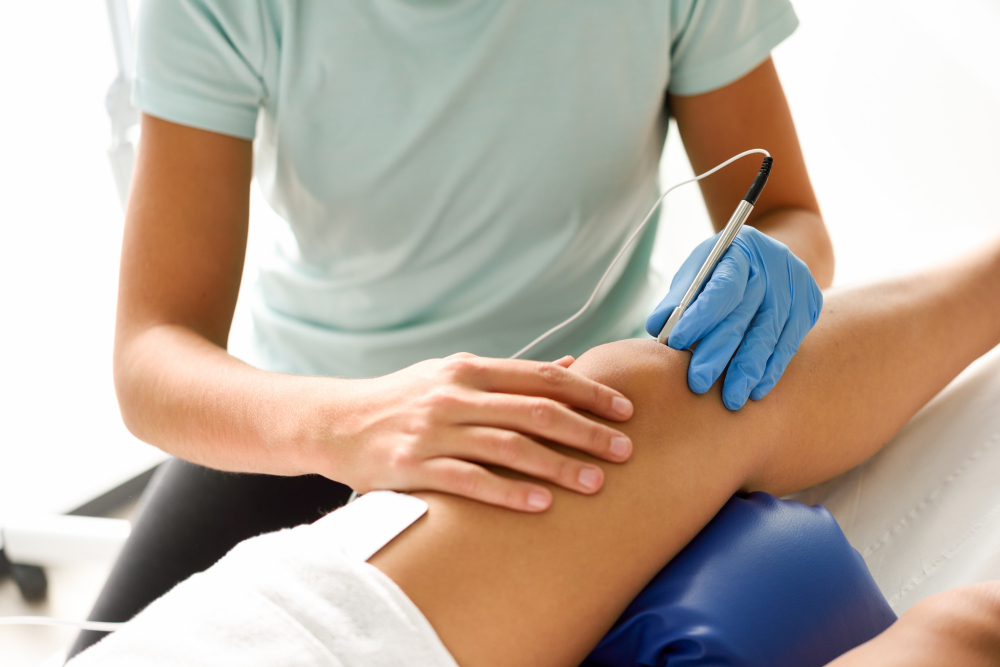
Conditions Treated With Knee Arthroscopy
Meniscus Tears
The most common reason for knee arthroscopy. The meniscus is a C-shaped cartilage cushion between the thigh and shin bones. Tears happen from twisting injuries, deep squats, sports collisions or with age-related degeneration.
Cartilage (Articular) Damage
Loose cartilage flaps, chondral defects, osteochondral lesions. Can be trimmed, drilled (microfracture) or treated with cartilage restoration techniques.
Loose Bodies
Free-floating bone or cartilage fragments inside the knee that cause locking and catching. Removed easily with arthroscopy.
Patellar (Kneecap) Problems
Recurrent kneecap dislocation, patellar instability, chondromalacia patellae. Treated with arthroscopic procedures including lateral release, MPFL reconstruction or cartilage interventions.
Synovitis
Inflammation of the joint lining. Synovial biopsy or partial synovectomy can be done arthroscopically.
Arthrofibrosis (Stiff Knee)
After previous surgery or trauma, scar tissue in the knee can severely limit movement. Arthroscopic adhesiolysis releases the scar tissue.
Knee Infections
Acute septic arthritis can be washed out arthroscopically - a procedure called arthroscopic lavage.
Meniscus Repair vs Meniscectomy - The Modern Approach
This is the most important decision in knee arthroscopy. Twenty years ago, surgeons routinely trimmed any torn meniscus (meniscectomy). Today we know that even partial meniscus removal increases the risk of knee arthritis significantly - by some estimates, advancing the arthritis timeline by 10 to 15 years.
Meniscus Repair (Stitching the Torn Meniscus Back Together)
Preferred whenever possible. Indicated for tears in the outer one-third of the meniscus (where blood supply allows healing), longitudinal tears, traumatic tears in young patients.
- Recovery: 6 to 12 weeks of restricted weight-bearing
- Long-term arthritis risk: significantly lower than meniscectomy
- Best option for patients under 50 with traumatic tears
Partial Meniscectomy (Trimming the Torn Portion)
Used when the tear is in the inner two-thirds of the meniscus (poor blood supply, won't heal) or when the tear is too degenerate to repair.
- Recovery: 2 to 4 weeks
- Long-term arthritis risk: noticeably increased
- Reserved for tears that cannot be repaired
Conservative (No Surgery) Treatment for Meniscus Tears
Many degenerative meniscus tears in patients above 50 do well with physiotherapy, injections and activity modification. Surgery is not always the answer. We recommend it only when mechanical symptoms (locking, catching, persistent giving way) are present.
How Knee Arthroscopy Is Performed
Under regional or general anesthesia. The patient lies supine. The leg is positioned in a special holder. The surgical team prepares and drapes the knee.
Three small incisions of 5 to 8 millimetres are made at the front of the knee. Sterile fluid is pumped in to expand the joint and improve visibility. The arthroscope is inserted through one portal. Instruments enter through the others. The surgeon watches a high-definition monitor and works inside the knee with millimetre precision.
Diagnostic arthroscopy is the first step - every part of the joint is inspected. Then the specific procedure is performed: meniscus repair, meniscus trimming, cartilage debridement, loose body removal, or whatever else is needed. After the procedure, the saline is drained, instruments removed and incisions closed with one or two stitches each.
Total surgery time: 30 to 90 minutes depending on what is being done.
Pricing for Knee Arthroscopy in Pune
- Diagnostic knee arthroscopy: Rs. 60,000 to Rs. 90,000
- Meniscus trimming: Rs. 70,000 to Rs. 1.1 lakh
- Meniscus repair: Rs. 90,000 to Rs. 1.4 lakh
- Cartilage procedures (microfracture, debridement): Rs. 80,000 to Rs. 1.3 lakh
- Patellar realignment, MPFL reconstruction: Rs. 1 to 1.5 lakh
- Loose body removal: Rs. 60,000 to Rs. 90,000
- Combined procedures (meniscus + cartilage + ligament work): Rs. 1.4 to 2 lakh
Most arthroscopies are day-care or 24-hour stay procedures. Insurance plans typically cover knee arthroscopy on cashless basis.
Recovery After Knee Arthroscopy
After Diagnostic Arthroscopy or Loose Body Removal
Walking immediately. Return to desk work in 3 to 7 days. Full activity in 2 to 4 weeks.
After Meniscus Trimming
Walking with stick for 1 to 2 weeks. Return to desk work in 1 to 2 weeks. Full sport return in 4 to 6 weeks.
After Meniscus Repair
Restricted weight-bearing for 4 to 6 weeks. Knee bending limited initially. Return to desk work in 2 to 4 weeks. Full sport return in 4 to 6 months.
After Cartilage Procedures
Variable - depends on the specific procedure. Microfracture requires 6 to 8 weeks of restricted weight-bearing. Cartilage transplant procedures may need 3 to 6 months of restricted activity.